
BIOLOGY / CHAPTER 01
The Cell: Structure, Organelles and Division
Chapter contents
Contents
Discovery of the cell and the cell theory
Cell size, shape and number
Prokaryotic cells
Eukaryotic cells: an overview; plant and animal cells compared
Cell wall
Plasma membrane and transport
The endomembrane system: ER, Golgi, lysosomes, vacuoles
Mitochondria
Plastids
Ribosomes
Cytoskeleton; cilia, flagella and centrioles
The nucleus and chromosomes
Microbodies
Enzymes and the chemistry inside the cell
The cell cycle and its regulation
Mitosis
Meiosis
Mitosis and meiosis compared; significance of cell division
Applied notes
Glossary ·
Revision points
1. Discovery of the Cell and the Cell Theory
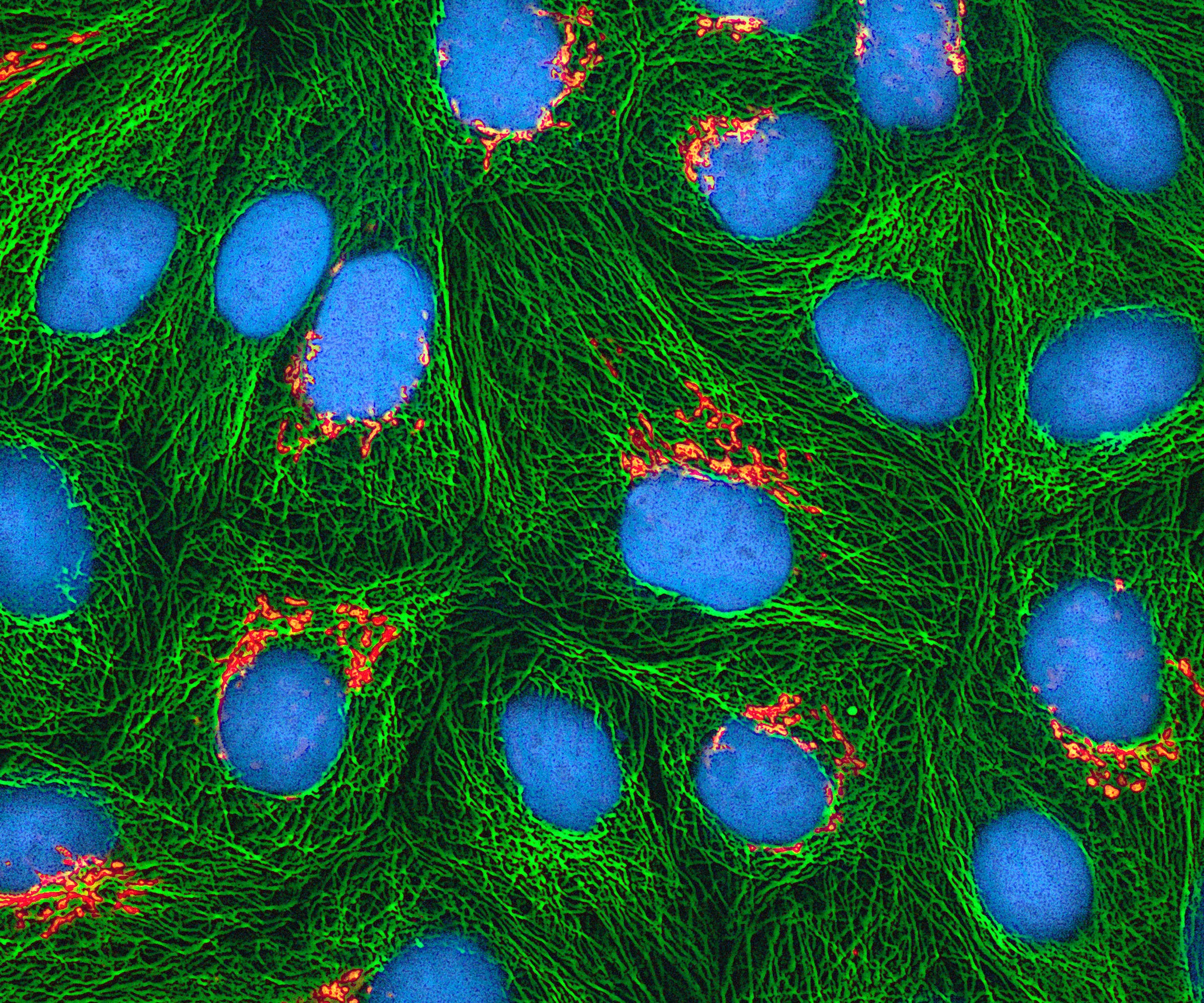
1.1 Historical landmarks
Scientist | Year | Contribution |
|---|---|---|
Robert Hooke | 1665 | Examined a thin slice of cork under a self-made microscope and saw honeycomb-like compartments, which he called "cells" (Latin cellula, a little room). He in fact saw only the dead cell walls. Described in Micrographia. |
Anton von Leeuwenhoek | 1674 | First to observe and describe living cells |
Robert Brown | 1831 | Discovered the nucleus in orchid cells. |
Dujardin / Purkinje | 1835 / 1839 | Dujardin described the living substance as "sarcode"; Purkinje coined the term "protoplasm". |
M.J. Schleiden | 1838 | A German botanist - concluded after studying many plants that all plants are composed of cells. |
Theodor Schwann | 1839 | A British zoologist: studied animal cells, reported that cells have a thin outer layer (the plasma membrane), that plant cells have a cell wall, and concluded that the bodies of animals and plants are composed of cells and their products. |
Rudolf Virchow | 1855 | Explained that cells divide and new cells are formed from pre-existing cells, "Omnis cellula e cellula". This completed the cell theory. |
Camillo Golgi; Altmann; Benda; Palade; de Duve | 1890–1955 | Discovery of the |
1.2 The cell theory
Contributions to cell theory
As formulated by Schleiden and Schwann and completed by Virchow, the cell theory states:
All living organisms are composed of cells and the products of cells.
The cell is the basic structural and functional unit of life: the smallest unit capable of independent existence. (70th BPSC CCE)
All cells arise from pre-existing cells by division.
Viruses, viroids and prions are acellular; they lack a cellular organisation and reproduce only inside a host cell. They are therefore not covered by the theory.
Coenocytic and syncytial structures (Rhizopus, Vaucheria, striated muscle fibres and the liquid endosperm of coconut) contain many nuclei in a continuous mass of cytoplasm without cell boundaries.
Mature mammalian red blood cells and sieve tube elements lack a nucleus; mature RBCs also lack most organelles.
Bacteria and cyanobacteria lack a well-defined nucleus and membrane-bound organelles, so the "typical cell" described by the theory does not fit them.
2. Cell Size, Shape and Number
Cell Type | Measurement | Key Feature |
|---|---|---|
Mycoplasma (PPLO) | 0.3 μm | Smallest known living cell |
Human Red Blood Cell | 7 μm | Standard human blood cell size |
Ostrich egg | 15 to 18 cm | Largest isolated single cell |
Nerve cell (Neuron) | Up to 1 metre | Longest cell in human body |
Shape varies with function: Discoidal (RBC), spherical, polygonal, columnar, spindle-shaped (muscle), branched with long processes (nerve).
Number: Organisms may be
1. Unicellular (Amoeba, bacteria, Chlamydomonas) or
2. multicellular.Cell size is limited by the surface area to volume ratio and by the capacity of the nucleus to control the cytoplasm.
3. Prokaryotic Cells
Represented by bacteria, cyanobacteria (blue-green algae), mycoplasma and PPLO. They multiply rapidly and are generally smaller than eukaryotic cells.
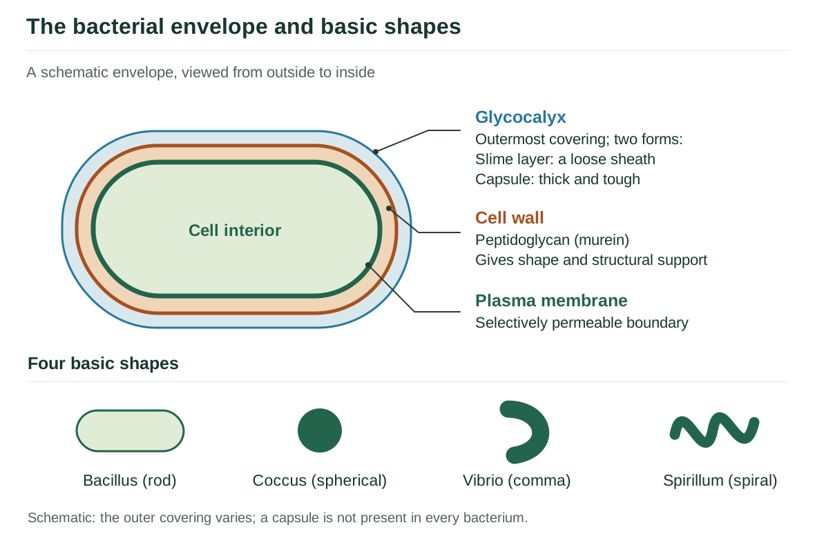
The four basic bacterial shapes are:
1. Bacillus (rod),
2. Coccus (spherical),
3. Vibrio (comma) and
4. Spirillum (spiral).
3.1 The cell envelope
The three-layered prokaryotic envelope
 Read the explanation
 Most prokaryotes have a chemically complex three-layered envelope, tightly bound and functioning as a unit:
Glycocalyx- the outermost layer. It may be a loose sheath called a slime layer, or a thick and tough capsule. Capsules protect against phagocytosis and desiccation, and contribute to virulence.
Cell wall: made of peptidoglycan (murein); determines the shape of the cell and provides strong structural support to prevent bursting or collapse.
Plasma membrane: selectively permeable; structurally similar to that of eukaryotes.
Gram staining (Christian Gram): bacteria that retain the stain are Gram-positive; those that do not are Gram-negative. The difference lies in the thickness of the peptidoglycan layer and the presence of an outer lipopolysaccharide membrane in Gram-negative cells, and it has major consequences for antibiotic sensitivity.
4. Eukaryotic Cells
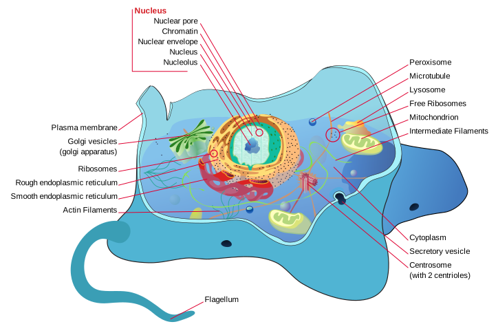
Found in protists, plants, animals and fungi. Their defining features are a true nucleus bounded by a nuclear envelope, and membrane-bound organelles, which permit compartmentalisation of function.
4.1 Prokaryotic and eukaryotic cells compared
Feature | Prokaryotic | Eukaryotic |
|---|---|---|
Size | Small, 1–10 µm | Larger, 5–100 µm |
Nucleus | Absent: nucleoid without a membrane | Present, with a nuclear envelope and nucleolus |
DNA | Single circular, naked, without histones | Linear, associated with histones, in several chromosomes |
Membrane-bound organelles | Absent | Present: ER, Golgi, mitochondria, lysosomes, plastids |
Ribosomes | 70S (50S + 30S) | 80S (60S + 40S) in the cytoplasm; 70S in mitochondria and chloroplasts |
Cell wall | Peptidoglycan | Cellulose in plants, chitin in fungi; absent in animals |
Cytoskeleton | Absent or rudimentary | Well developed |
Cell division | Binary fission; no spindle | Mitosis and meiosis; spindle formed |
Flagella | Made of flagellin; simple structure | Made of tubulin; 9 + 2 arrangement |
Examples | Bacteria, cyanobacteria, mycoplasma | Protists, fungi, plants, animals |
4.2 Plant and animal cells compared
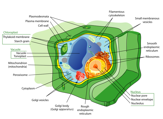
Feature | Plant cell | Animal cell |
|---|---|---|
Cell wall | Present : cellulose | Absent |
Plastids | Present | Absent |
Vacuole | A single large central vacuole, up to 90 per cent of cell volume | Small and numerous, or absent |
Centrosome / centrioles | Usually absent in higher plants | Present |
Lysosomes | Rare; the vacuole performs the function | Present and prominent |
Shape | Fixed, generally rectangular | Variable, generally rounded |
Cytokinesis | By cell plate formation | By cleavage furrow |
Reserve food | Starch | Glycogen |
Plasmodesmata | Present | Absent; gap junctions instead |
5. Cell Wall
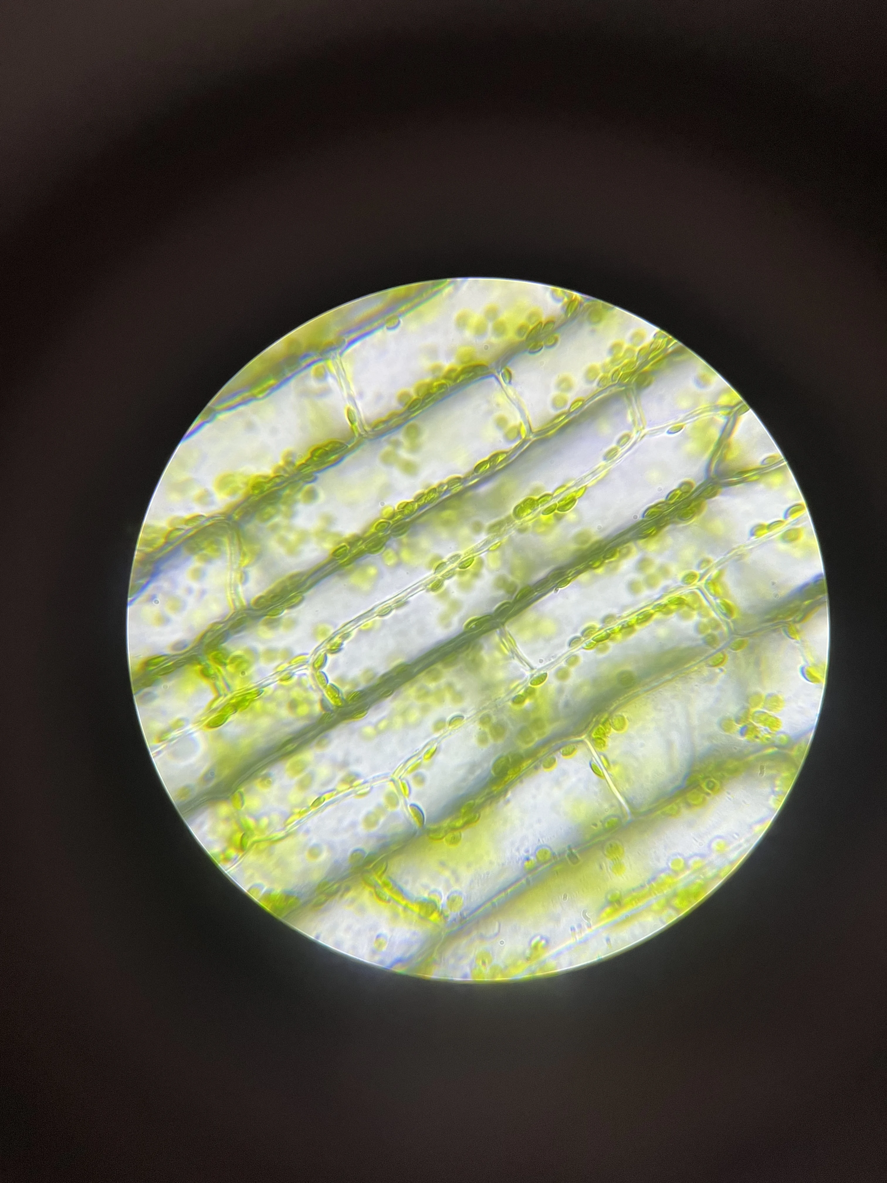
General Characteristics
Cell wall: A non-living rigid structure forming an outer covering for the plasma membrane in fungi and plants.
Functions: Protects cell, Maintains shape, and Prevents osmotic lysis.
Animal cells: Completely lack a cell wall.
Cell Wall Composition by Organism Group
Organism Group | Main Cell Wall Material | Structural Details |
|---|---|---|
Plants | Cellulose | Primary wall: cellulose, hemicellulose, pectin. Middle lamella: calcium/magnesium pectate. Secondary wall: lignin or suberin. |
Fungi | Chitin | Polymer of N-acetylglucosamine; cellulose is absent. |
Bacteria | Peptidoglycan (Murein) | Polysaccharides cross-linked by short peptides. Thick in Gram-positive, thin in Gram-negative. |
Algae | Cellulose and Galactans | Contains cellulose, galactans, mannans, and minerals like calcium carbonate. |
Animals | Absent | No cell wall present. |
6. Plasma Membrane and Transport
6.1 Chemical composition
Membrane composition: composed primarily of lipids (chiefly phosphoglycerides) and proteins (71st BPSC CCE).
The ratio of protein to lipid varies greatly: human RBC membrane has about 52 per cent protein and 40 per cent lipid.
6.2 Models of membrane structure
Fluid Mosaic Model: Universally accepted model, proposed by Singer and Nicolson in 1972.
Structure: Proteins float within a continuous two-dimensional liquid phospholipid bilayer.
6.3 Transport across the membrane
Membrane transport: direction and ATP
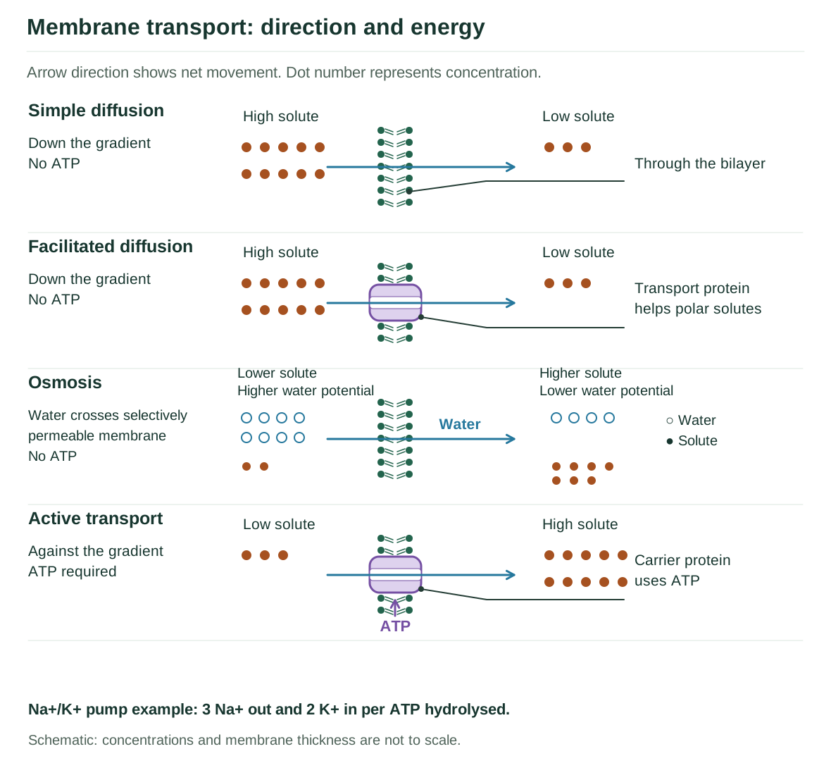 Read the explanation
 Mode | Energy | Description |
|---|---|---|
Simple diffusion | None | Passive movement down a concentration gradient. Neutral solutes cross freely; non-polar (lipid-soluble) molecules pass most readily. |
Osmosis | None | Diffusion of water across a semi-permeable membrane from a region of higher to lower water potential. |
Facilitated diffusion | None | Polar molecules, which cannot cross the non-polar interior unaided, are carried by membrane transport proteins, still down the gradient. |
Active transport | ATP required | Carrier proteins pump molecules against the concentration gradient. Example: the sodium–potassium pump, which moves 3 Na+ out and 2 K+ in per ATP hydrolysed. |
Bulk transport | ATP required | Endocytosis: phagocytosis ("cell eating", of solid particles), pinocytosis ("cell drinking", of fluids) and receptor-mediated endocytosis. Exocytosis: expulsion of material in vesicles that fuse with the plasma membrane. |
Osmosis is a passive transport process where water moves from lower solute concentration to higher solute concentration across a selectively permeable membrane.
7. The Endomembrane System
The organelles whose functions are coordinated are considered together as the endomembrane system:
1. Endoplasmic reticulum,
2. Golgi complex,
3. Lysosomes and
4. Vacuoles.
5. Mitochondria,
6. Chloroplasts and
7. Peroxisomes are excluded because their functions are not coordinated with these.
Endomembrane organelles and their coordinated roles
7.1 Endoplasmic reticulum
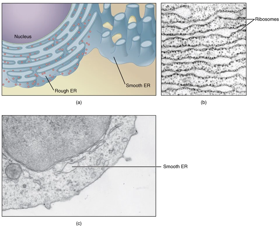
The endoplasmic reticulum (ER) is like a transport system inside a cell. It is a network of tiny tubes that helps make and move proteins and fats around the cell.
There are two types:
Rough ER: Has ribosomes on it and makes proteins.
Smooth ER: Has no ribosomes and makes Lipid and steroid synthesis, drug detoxification: helps remove harmful substances.
7.2 Golgi apparatus
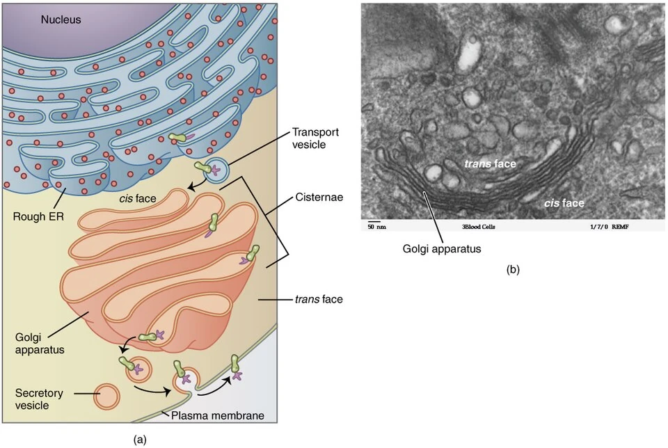
Discovered by Camillo Golgi in 1898 in nerve cells; also called the dictyosome in plants.
Consists of many flat, disc-shaped sacs or cisternae, 0.5–1.0 µm in diameter, stacked parallel and concentrically arranged near the nucleus.
Functions:
The principal site of packaging and dispatch of materials;
Modification of proteins, especially glycosylation;
Formation of glycolipids and glycoproteins;
Formation of lysosomes; and,
In plant cells, synthesis of the polysaccharides of the cell wall.
Aptly called the "post office" or the "traffic police" of the cell.
7.3 Lysosomes
Discovered by Christian de Duve; membrane-bound vesicular structures formed by the packaging activity of the Golgi apparatus.
Contain almost fifty hydrolytic enzymes (lipases, proteases, carbohydrases and nucleases) all of which are optimally active at acidic pH (about 5), maintained by proton pumps in the lysosomal membrane.
Types: primary (freshly formed, enzymes inactive), secondary or digestive vacuole (formed by fusion with a food vacuole), residual body (containing undigested material), and autophagic vacuole (digesting the cell's own worn-out organelles).
Enzyme nature: Lysosomes contain hydrolytic enzymes; most enzymes are proteins. (67th BPSC CCE)
Functions:
Intracellular digestion;
Destruction of foreign material;
Autophagy and Autolysis: the self-destruction of the cell, which has earned lysosomes the name "suicide bags of the cell".
Also important in metamorphosis (resorption of the tadpole's tail).
7.4 Vacuoles
A vacuole is like a storage bag inside a cell.
A membrane-bound space in the cytoplasm containing water, sap, excretory products and other materials not useful to the cell.
Bounded by a single membrane called the tonoplast.
In the plant cell the vacuole may occupy up to 90 per cent of the cell volume and is responsible for turgidity.
The tonoplast facilitates the transport of ions against the concentration gradient, so the vacuole may hold a higher concentration of ions than the cytoplasm.
In Amoeba the contractile vacuole is important in osmoregulation and excretion; many protists have food vacuoles formed by engulfing food particles.
8. Mitochondria: The Powerhouse of the cell
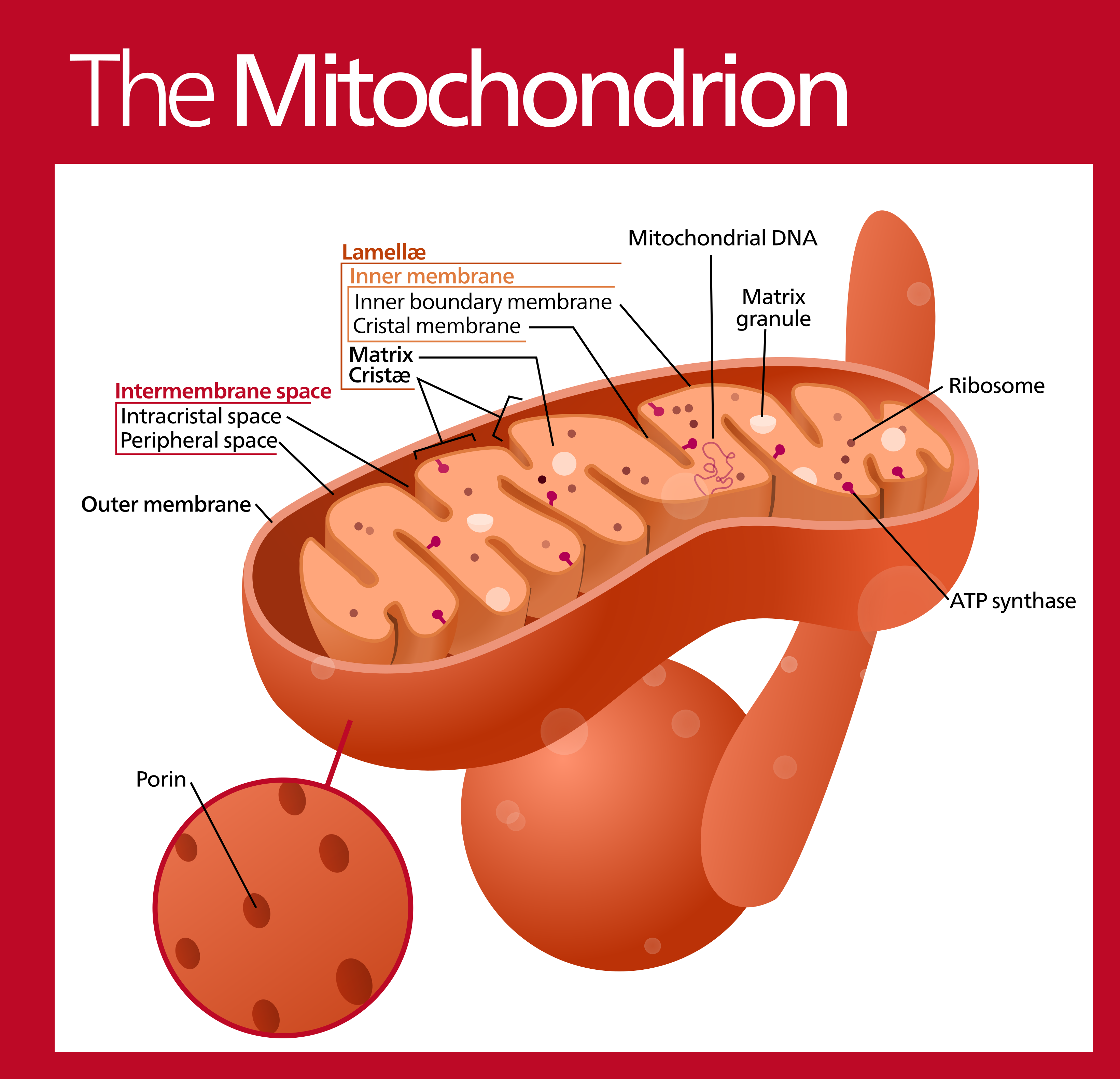
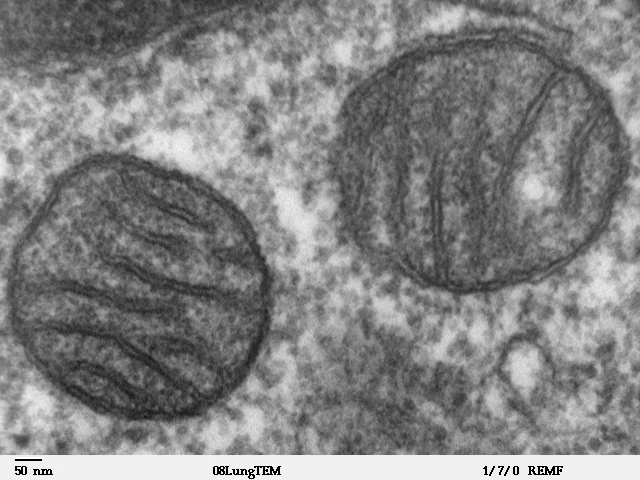
First observed by Richard Altmann (1890) as "bioblasts" and named mitochondria by Carl Benda (1897).
Shape and size: usually cylindrical or sausage-shaped
Number varies with the physiological activity of the cell.
Active cells: High numbers in muscle and nerve cells for energy demands.
Structure: A double membrane:
The outer membrane is smooth and forms a continuous limiting boundary; it is permeable to small molecules (porins).
The inner membrane forms a number of infoldings called cristae towards the matrix, which greatly increase the surface area. It is selectively permeable.
The two membranes divide the organelle into two aqueous compartments, the outer (intermembrane) compartment and the inner compartment or matrix.
The inner membrane bears stalked particles: F0-F1 particles, also called oxysomes or elementary particles: which carry out ATP synthesis.
Matrix contents:
A single circular DNA molecule,
A few RNA molecules,
70S ribosomes and
The components required to synthesise proteins.
Mitochondria therefore divide by fission and are described as semi-autonomous organelles.
Function:
The site of aerobic respiration- the Krebs cycle in the matrix and the electron transport chain and oxidative phosphorylation on the inner membrane.
They produce cellular energy in the form of ATP, and are therefore called the "powerhouses of the cell".
9. Plastid Family and Chloroplast
Found in all plant cells and in euglenoids. They bear specific pigments and are classified into three types.
Type | Description |
|---|---|
Chloroplasts | Contain chlorophyll and carotenoid pigments and trap light energy for photosynthesis. In higher plants they are lens-shaped, oval or ribbon-like, 5–10 µm long and 2–4 µm wide; Their number varies from one per cell in Chlamydomonas to 20–40 in a mesophyll cell. |
Chromoplasts | Contain carotenoids- carotene, xanthophylls : giving the plant part a yellow, orange or red colour (Fruits, Petals). |
Leucoplasts | Colourless plastids of varied shape and size, storing nutrients: 1. Amyloplasts (starch), |
Structure of the chloroplast
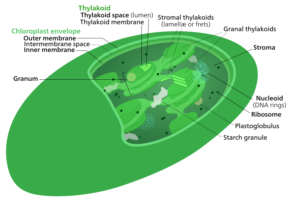
Bounded by a double membrane; the space inside is the stroma.
The stroma contains a number of organised flattened membranous sacs called thylakoids. Thylakoids are arranged in stacks called grana (singular granum), and flat membranous tubules called stroma lamellae connect the thylakoids of different grana.
The space inside a thylakoid is the lumen.
The light reactions occur on the thylakoid membranes;
The dark reactions (carbon fixation) occur in the stroma.
The stroma also contains circular DNA, 70S ribosomes and enzymes - so chloroplasts too are semi-autonomous.
Semi-autonomous organelles: Why DNA and RNA enable self-replication
Why organelles are semi-autonomous
Replication: Mitochondria and chloroplasts duplicate independently via binary fission.
Dependence: Rely on nuclear DNA for most structural proteins and enzymes.
The presence of DNA alone does not allow self-replication; requires both DNA and RNA with 70S ribosomes. (68th BPSC CCE)
THE ENDOSYMBIOTIC THEORY (Lynn Margulis)
Mitochondria and chloroplasts both possess a double membrane, their own circular DNA, 70S ribosomes, and the ability to divide independently by fission: all features of prokaryotes.
The endosymbiotic theory proposes that they originated as free-living prokaryotes (an aerobic bacterium and a cyanobacterium respectively) that were engulfed by an ancestral eukaryotic cell and retained in a mutually beneficial relationship. This is the standard explanation for why they are semi-autonomous.
10. Ribosomes: Protein Factories
Ribosome subunits and protein destination
80S (60S + 40S)
70S (50S + 30S)
free in the cytoplasm
Rough Endoplasmic Reticulum
First observed under the electron microscope by George Palade (1953).
Granular structures, not surrounded by any membrane - the only major organelle without a membrane.
Composed of ribosomal RNA (rRNA) and proteins, in two unequal subunits that associate only in the presence of Mg2+.
Ribosome types:
80S (60S + 40S) in eukaryotic cytoplasm;
70S (50S + 30S) in prokaryotes, mitochondria, and chloroplasts.
"S" is the Svedberg unit, a measure of the sedimentation coefficient, which depends on both size and shape.
Found free in the cytoplasm (proteins for use within the cell) or attached to the Rough Endoplasmic Reticulum (proteins for export or for membranes).
Several ribosomes translating a single mRNA form a polysome.
Function: The site of protein synthesis : the "Protein Factories" of the cell.
11. Cytoskeleton, Cilia, Flagella and Centrioles
11.1 Cytoskeleton
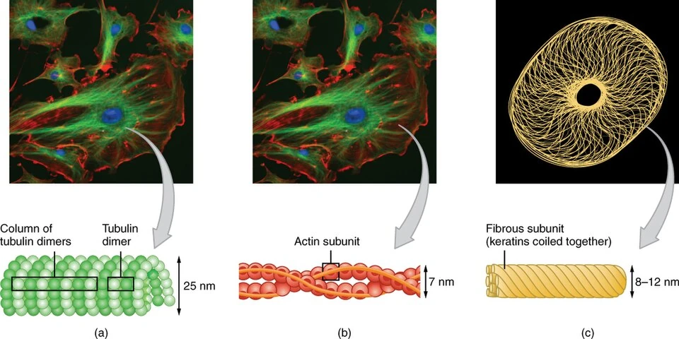
The cytoskeleton is a network of protein fibres inside the cell. It gives the cell shape, support and movement., of three kinds:
Microtubules : Hollow tubes made of tubulin. They help form spindle fibres, cilia, flagella and centrioles.
Microfilaments: Very thin fibres made of actin. They help in cell contraction and movement of cytoplasm.
Intermediate filaments: Provide tensile strength. Give the cell strength and support
11.2 Cilia and flagella
Cilia and flagella are hair-like structures that come out from the cell.
Cilia are small and numerous and work like oars, moving the cell or the surrounding fluid.
Flagella are longer and fewer and are responsible for cell movement.
Both are covered with the plasma membrane. The core, called the axoneme, possesses a number of microtubules running parallel to the long axis.
11.3 Centrosome and centrioles
TThe centrosome is an organelle found mainly in animal cells. It usually contains two centrioles.
The two centrioles are arranged at right angles (90°) to each other.
Functions:T
The centrioles form the basal body of cilia and flagella, and
Give rise to the spindle apparatus during cell division in animal cells.
12. The Nucleus and Chromosomes
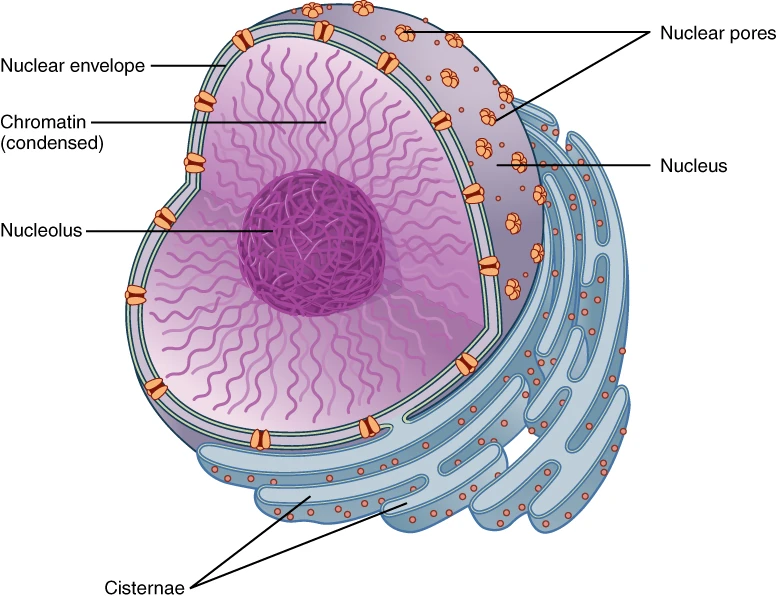
12.1 Structure and Functions
Control centre: Directing growth, metabolism, and division.
First described by Robert Brown (1831); the stainable material was called chromatin by Flemming.
Biological membranes: Nuclear membrane and plasma membrane are composed of proteins and lipids. (71st BPSC CCE)
Number of nuclei: usually one; some cells are anucleate (mature mammalian RBC, sieve tube cells) and some multinucleate (Rhizopus, striated muscle fibres).
Components of the nucleus
Component | Structure | Primary Function |
|---|---|---|
Nuclear membrane | Double membrane penetrated by nuclear pores | Regulates transport of RNA and proteins |
Nucleoplasm | Enclosed fluid matrix | Houses chromatin, nucleolus, enzymes, and nucleotides |
Nucleolus | Dense, Non-membrane-bound body | Synthesises ribosomal RNA (rRNA) |
Chromatin | DNA wrapped around basic histone proteins | Condenses into chromosomes during division |
12.2 Chromatin and chromosomes
From nucleus to DNA packaging
Histone octamer: 2 × H2A + 2 × H2B + 2 × H3 + 2 × H4. Histones are positively charged.
About 200 base pairs of DNA are associated with each nucleosome.
Chromatin is an material inside nucleus (an interphase network of nucleoprotein fibres).
It’s made up of: DNA, histones, non-histone proteins and RNA.
When the cell is not dividing (Interphase), DNA is present as a loose network called chromatin.
During division it condenses into chromosomes.
Chromatin = Loose DNA
Chromosome = Condensed/tightly packed DNATypes of Chromatin:
Euchromatin: Loosely packed, lightly stained, transcriptionally active. Heterochromatin: Densely packed, darkly stained, transcriptionally inactive.Nucleosome model: A nucleosome is the basic repeating unit of chromatin.
Think of DNA as a long thread wrapped around a spool.
DNA is negatively charged.
Histone proteins are positively charged.
DNA wraps around a group of 8 histone proteins, called a histone octamer.
The octamer contains:
2 × H2A
2 × H2B
2 × H3
2 × H4
About 200 base pairs of DNA are associated with each nucleosome.
Parts and types of a chromosome
Every chromosome has a primary constriction or centromere, on the sides of which are disc-shaped structures called kinetochores, to which the spindle fibres attach. Some chromosomes have a secondary constriction with a small terminal fragment called the satellite.
Type | Position of the centromere and shape at anaphase |
|---|---|
Metacentric | Centromere in the middle; two equal arms; appears V-shaped at anaphase. |
Sub-metacentric | Centromere slightly away from the middle; one arm shorter than the other; appears L-shaped. |
Acrocentric | Centromere situated close to one end; one very short and one very long arm; appears J-shaped. Often bears a satellite. |
Telocentric | Centromere at the terminal end; a single arm; appears I- or rod-shaped. Not found in humans. |
13. Microbodies
Microbodies: three roles
Membrane-bound minute vesicles containing various enzymes, present in both plant and animal cells.
Peroxisomes:
Contain oxidases and catalase;
carry out oxidation reactions producing hydrogen peroxide, which catalase then breaks down.
Involved in photorespiration in plants and in the detoxification of alcohol in liver cells.
Glyoxysomes- Found in germinating fatty seeds; site of the glyoxylate cycle, converting stored fats into carbohydrates.
Sphaerosomes- Associated with the synthesis and storage of lipids in plant cells.
14. Enzymes and the chemistry inside the cell
14.1 What an enzyme is, why it is called a biocatalyst, and why almost all of them are proteins
Biocatalyst: Produced by living organisms to speed up reactions without being consumed.
Lowers activation energy so metabolic reactions occur rapidly at body temperature.
Chemically, almost all enzymes are proteins made of long amino acid chains. (67th BPSC Re-Exam)
14.2 Substrate, active site, and models of enzyme action
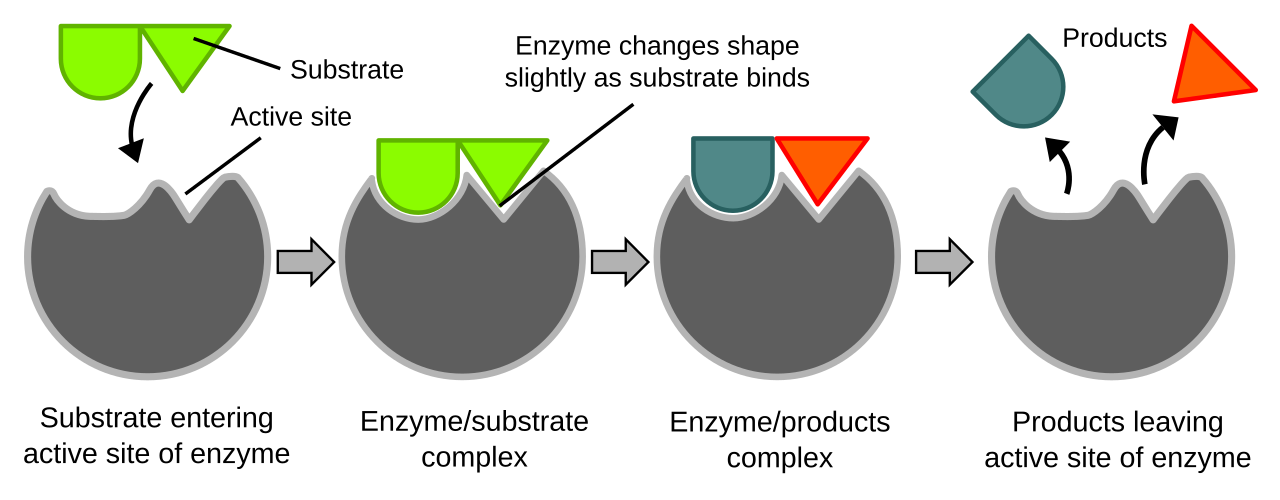
Mechanism of Action
Substrate: Reactant molecule acted upon by an enzyme.
Active Site: Specific enzyme region where substrate binds to form temporary enzyme-substrate complex.
Models of Enzyme Action
Model | Proposed By | Key Concept" |
|---|---|---|
Lock-and-Key Model | Emil Fischer (1894) | Active site is rigid and fits only a matching substrate shape. |
Induced-Fit Model | Daniel Koshland (1958) | Active site is flexible and alters shape slightly for tight fit. |
14.3 Temperature and pH: factors affecting enzyme activity
Temperature Effects
Optimum Temperature: 37 °C (98.6 °F) for most human enzymes.
Cold Temperatures: Temporarily deactivate enzyme activity.
High Temperatures: Permanently alter protein structure causing denaturation.
Optimum pH for Digestive Enzymes
Enzyme | Location | Optimum pH | Environment |
|---|---|---|---|
Pepsin | Stomach | 1.5 to 2.0 | Strongly acidic |
Salivary Amylase | Mouth | 6.8 | Weakly acidic / neutral |
Trypsin | Small Intestine | 8.0 | Alkaline |
Extreme pH changes break ionic and hydrogen bonds, permanently altering active site shape and causing denaturation.
14.4 Ribozymes and the RNA exception
Discovery of Non-Protein Catalysts
1980s: Thomas Cech and Sidney Altman discover catalytic RNA molecules called ribozymes.
Ribozyme Functions
Composed of RNA rather than amino acids or proteins.
Catalyze joining of amino acids during protein synthesis in ribosomes.
15. The Cell Cycle
The cell cycle is the series of steps a cell goes through to grow, copy its DNA, and divide into two new cells (daughter cells).
It has two broad phases:
Interphase: Cell grows and prepare for division.
M phase: Cell divides
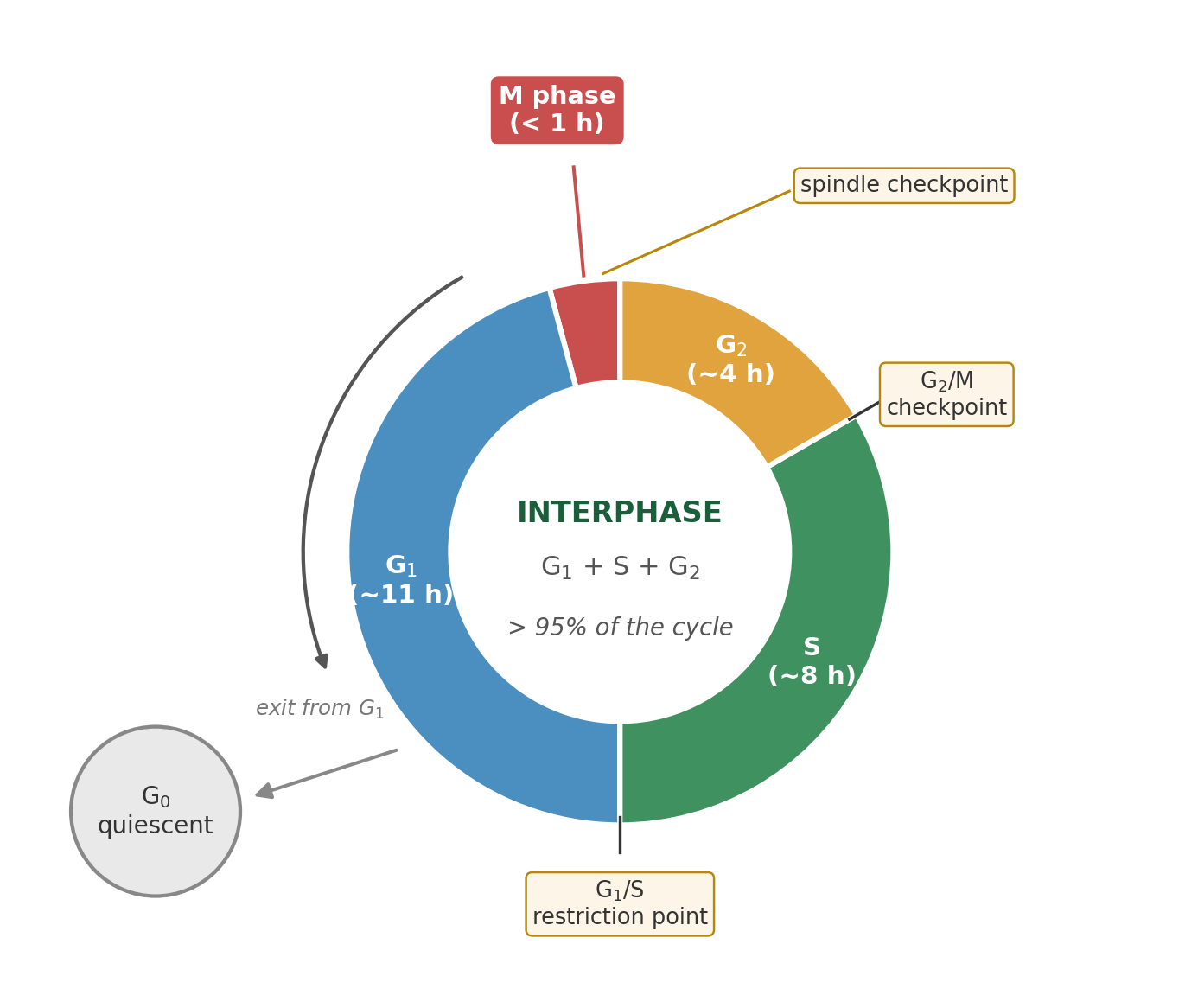
The eukaryotic cell cycle. Interphase (G1, S and G2) occupies more than 95 per cent of the cycle; the M phase lasts less than an hour in a typical human cell. Cells that stop dividing leave the cycle at G1 and enter the quiescent G0 stage.
15.1 Phases
The cell cycle
In S phase, the amount of DNA doubles (2C to 4C), but the total chromosome count stays 2n.
A typical human cell cycle lasts about 24 hours, of which interphase occupies more than 95 per cent; interphase is therefore not a "resting phase" but a period of intense preparatory activity.
Phase | Duration | Events |
|---|---|---|
G1 (Gap 1) | About 11 hours | The cell is metabolically active and grows continuously but does not replicate its DNA. Synthesis of RNA, proteins and organelles. The G1 restriction point lies at the end of this phase. |
S (Synthesis) | About 8 hours | DNA replication takes place: DNA is copied. The amount of DNA doubles (2C → 4C), but the number of chromosomes does not increase- each chromosome now has two chromatids. In animal cells the centriole duplicates in the cytoplasm during this phase. |
G2 (Gap 2) | About 4 hours | Proteins are synthesised in preparation for mitosis; the cell continues to grow. The G2/M checkpoint lies at the end. |
M (Mitotic) | Less than 1 hour | The actual division: karyokinesis (nuclear division) followed by cytokinesis (cytoplasmic division). |
In S phase, the amount of DNA doubles (2C to 4C), but the total chromosome count stays 2n.
Points to note:
A typical human cell cycle lasts about 24 hours, of which interphase occupies more than 95 per cent; interphase is therefore not a "resting phase" but a period of intense preparatory activity.
In yeast the cycle is completed in about 90 minutes.
15.2 The G0 or quiescent stage
Some cells stop dividing and leave the cell cycle after G1. These cells are still metabolically active, but they do not normally divide.
Heart cells → usually permanently in G0
Nerve cells → usually permanently in G0
Liver cells → can return to the cell cycle when needed
G0 = Cell is alive and working, but not dividing.
15.3 Regulation of the cell cycle
The cell cycle is controlled by proteins called:
Cyclins
CDKs (Cyclin-dependent kinases)
Think of them as the control system/traffic lights of the cell cycle.
A CDK is inactive on its own and becomes active only when bound to its cyclin; the concentration of each cyclin rises and falls at a specific point in the cycle.
15.4 Checkpoints
Checkpoints make sure everything is correct before the cell moves to the next stage.
1. G1/S checkpoint
Is the cell big enough?
Are there enough nutrients?
Is the DNA undamaged?
2. G2/M checkpoint
Has DNA been copied correctly?
3. M checkpoint (spindle checkpoint)
Are all chromosomes properly attached to spindle fibres?
Why are checkpoints important?
They prevent cells with serious problems from dividing.
Cancer: If these controls fail because of mutations in genes such as p53, cells may divide uncontrollably, which can lead to cancer.
REMEMBER:
Cell Cycle = Grow → Copy DNA → Prepare → Divide
Phases:
G1 = Grow
S = DNA Synthesis
G2 = Prepare
M = Divide
G0 = Not dividing
Checkpoints = Safety checks
Checkpoint failure → Uncontrolled cell division → Cancer
16. Mitosis
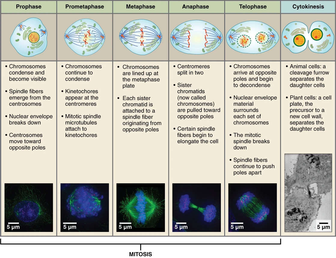
Produce two genetically identical diploid (2n) daughter cells.
Also called equational division, because the daughter cells receive the same number of chromosomes as the parent.
It occurs in somatic cells (and in the diploid cells of plants, and in haploid cells of some organisms).
The division occurs in phases:
karyokinesis (nuclear division) followed by
cytokinesis (cytoplasmic division).
16.1 Karyokinesis (Nuclear division)
Stage | Events |
|---|---|
Prophase | Follows the S and G2 phases, in which the DNA has already replicated. DNA has already been copied during the S phase. Chromatin becomes tightly packed into visible chromosomes. Each chromosome has 2 sister chromatids joined at the centromere. The centrosomes move towards opposite sides of the cell. Spindle fibres begin to form. By the end, the nuclear envelope, nucleolus, Golgi body and ER disappear. |
Metaphase | The nuclear envelope is completely disintegrated, and the chromosomes are spread through the cytoplasm. Chromosomes are fully condensed. Chromosomes line up in the middle/equator of the cell. This is the best stage for studying chromosome morphology. Spindle fibres attach to the kinetochores on either side of the centromere. The chromosomes align at the equator, on the metaphase plate. |
Anaphase | The centromeres split. and The two sister chromatids separate. Each becoming a daughter chromosome. The chromosomes move towards opposite poles. |
Telophase | The chromosomes reaches at the poles and decondense, They become loose again and form chromatin. A nuclear envelope forms around each group. The nucleolus, Golgi complex and endoplasmic reticulum reappear. |
16.2 Cytokinesis (Cytoplasmic division)
Cytokinesis means division of the cytoplasm after the nucleus has divided.
In an animal cell:
A cleavage furrow (groove) appears in the cell membrane.
The furrow becomes deeper and deeper. It finally divides the cell into two daughter cells.
The furrow is made by a contractile ring of actin and myosin.
Division happens from outside → inside
In a plant cell:
The wall makes furrowing impossible.
Instead:A cell plate forms at the centre of the cell.
It grows outwards until it reaches the existing cell walls - that is, centrifugally.
The cell plate becomes the middle lamella between the two daughter cells.
Plant cell = Cell plate = Centre → Outside
16.3 Significance of mitosis
Produces daughter cells that are genetically identical to the parent cell - the basis of growth.
Maintains the same chromosome number generation after generation in somatic tissue.
Restores the nucleo-cytoplasmic ratio, which becomes disturbed as the cell grows.
Enables repair and replacement of worn-out cells - epidermis, gut lining, blood cells.
In plants, permits continuous growth throughout life through meristematic tissue.
The basis of asexual reproduction and of vegetative propagation.
17. Meiosis
Meiosis: one DNA copy, two divisions
A Meiosis is a special type of cell division that produces sex cells (gametes) such as sperm and eggs.
The main purpose is to reduce the chromosome number by half.
The most important idea:
1 diploid cell (2n) → 4 haploid cells (n)
There are 2 divisions: Meiosis I and Meiosis II.
But DNA is copied only once, before Meiosis I.
17.1 Meiosis I (= Reduction division)
Meiosis I is called the reduction division because the chromosome number is reduced by half.
Prophase I: the longest and most complex stage, divided into five sub-stages
Leptotene → Zygotene → Pachytene → Diplotene → Diakinesis
The important event in Prophase I is that homologous chromosomes pair up and exchange genetic material through crossing over.
The remaining stages of meiosis I
Metaphase I: Homologous pairs in the middle
Anaphase I: Homologues separate, sisters stay together.
Telophase I: 2 cells formed
Interkinesis: This is the short period between Meiosis I and Meiosis II, there is no replication of DNA (DNA was already copied before Meiosis I).
17.2 Meiosis II (= Equational division)
Similar to a normal mitosis and is equational.
Prophase II: the nuclear membrane disappears again; the chromosomes become compact.
Metaphase II: chromosomes align at the equator; spindle fibres attach to the kinetochores of the sister chromatids.
Anaphase II: the centromeres split and the sister chromatids separate, moving to opposite poles.
Telophase II: nuclear envelopes reform, chromosomes decondense, cytokinesis follows, producing four haploid daughter cells in a tetrad.
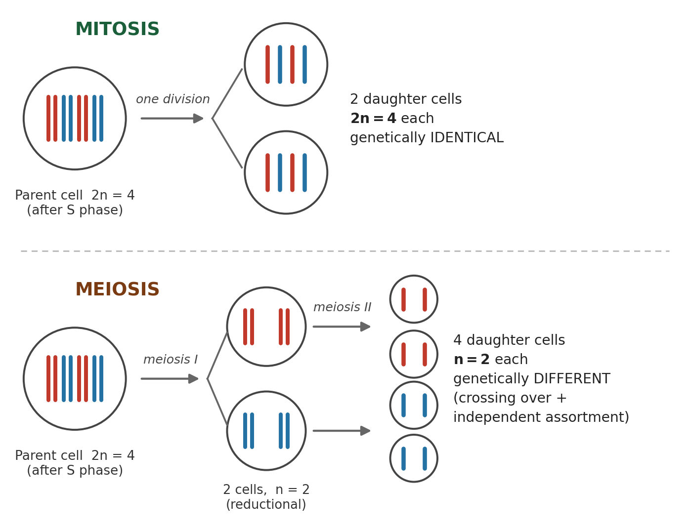
Outcome of mitosis and meiosis from a diploid parent cell (2n = 4). Mitosis yields two genetically identical diploid cells; meiosis, through one round of DNA replication followed by two divisions, yields four genetically distinct haploid cells.
17.3 Significance of meiosis
Maintains the chromosome number of a species across generations: gametes are haploid, so fertilisation restores the diploid number.
Introduces genetic variation through crossing over in pachytene and the independent assortment of maternal and paternal chromosomes at metaphase I.
This variation is the raw material of evolution and of natural selection.
Essential for sexual reproduction and gamete formation.
18. Mitosis and Meiosis Compared
Where each happens in the body, and the chromosome count after each
Body Location & Fertilization
Mitosis location: Somatic cells (skin, liver, bone) for growth, repair, and asexual reproduction.
Meiosis location: Germ cells (testes, ovaries, anthers, ovules) for gamete production.
Fertilization: Haploid sperm (23) fuses with haploid egg (23) to restore 46 chromosomes in zygote.
Feature | Mitosis | Meiosis |
|---|---|---|
Location | Somatic cells | Germ cells |
Divisions | 1 division | 2 divisions |
Daughter cells | 2 cells | 4 cells |
Genetic composition | Identical to parent | Genetically distinct |
Parent count | 46 chromosomes (23 pairs) | 46 chromosomes (23 pairs) |
Daughter count | 46 chromosomes (diploid / 2n) | 23 chromosomes (haploid / n) |
19. Applied Notes
Cellular failures and their outcomes
Cancer:
Arises from a failure of cell-cycle control: uncontrolled cell division (mitosis) and may spread by metastasis.
In tumour suppressor genes such as p53, the "guardian of the genome", are typically involved.
Apoptosis: Programmed cell death that removes unwanted/damaged cells.
Necrosis: Abnormal/pathological cell death.
Lysosomal storage disorders result from the deficiency of a single lysosomal enzyme, for example Tay-Sachs disease, Gaucher's disease and Pompe's disease.
Mitochondrial inheritance is maternal, i.e. Mitochondrial DNA is mainly inherited from the mother.
Non-disjunction: Failure of chromosomes to separate → abnormal chromosome number, giving rise to conditions such as Down syndrome (trisomy 21), Turner syndrome (45, XO) and Klinefelter syndrome (47, XXY).
Antibiotics and the 70S ribosome: Bacteria have 70S ribosomes; drugs like tetracycline and streptomycin inhibit bacterial protein synthesis.
Colchicine: Blocks tubulin/spindle formation, causing metaphase arrest; used in karyotyping and plant breeding.
20. Glossary
Term | Meaning |
|---|---|
Protoplasm | The living content of a cell: cytoplasm together with the nucleus. |
Cytosol | The aqueous ground substance of the cytoplasm, excluding the organelles. |
Nucleoid | The region of a prokaryotic cell containing the DNA, not bounded by a membrane. |
Mesosome | An infolding of the prokaryotic plasma membrane. |
Cristae | Infoldings of the inner mitochondrial membrane. |
Grana / thylakoid | Stacks of flattened membranous sacs in the chloroplast stroma / an individual sac. |
Semi-autonomous organelle | An organelle with its own DNA and ribosomes, able to divide: mitochondrion and plastid. |
Nucleosome | A histone octamer with about 200 bp of DNA wrapped around it: the repeating unit of chromatin. |
Kinetochore | The disc-shaped protein structure at the centromere to which spindle fibres attach. |
Synapsis / bivalent | The pairing of homologous chromosomes / the resulting paired structure of four chromatids. |
Chiasma | The visible X-shaped point of crossing over between non-sister chromatids. |
Syncytium | A multinucleate mass formed when karyokinesis is not followed by cytokinesis. |
G0 | The quiescent stage in which cells are metabolically active but not dividing. |
21. Rapid Revision: Facts and Numbers
Cell: The fundamental structural and functional unit of life discovered by Robert Hooke (1665) in cork, while Anton van Leeuwenhoek (1674) first observed living cells.
Cell Theory: Formulated by Schleiden (1838) and Schwann (1839), and completed by Rudolf Virchow (1855) with the principle *Omnis cellula e cellula* (cells arise from pre-existing cells).
Cell Membrane: A selectively permeable membrane described by Singer and Nicolson's Fluid Mosaic Model (1972), composed primarily of proteins and lipids.
Cell Wall: Absent in animals, but present in plants (cellulose), fungi (chitin), and bacteria (peptidoglycan).
Nucleus: The principal control centre discovered by Robert Brown (1831), containing chromatin (DNA and histones) and the nucleolus for rRNA synthesis.
Mitochondria: Known as the powerhouse of the cell, generating energy as ATP through aerobic respiration across inner membrane folds called cristae.
Chloroplast: A double-membrane plastid containing chlorophyll in thylakoid grana and stroma that carries out photosynthesis.
Semi-autonomous Organelles: Mitochondria and chloroplasts replicate independently because they contain their own circular DNA, RNA, and 70S ribosomes.
Organelles: Includes ribosomes (protein synthesis), Rough ER (protein processing), Smooth ER (lipid synthesis), Golgi body (packaging), lysosomes (hydrolytic digestion), and vacuoles (storage).
Enzymes: Biological catalysts that lower activation energy; almost all enzymes are proteins, except catalytic RNA molecules called ribozymes.
Mitosis: Equational division occurring in somatic cells, producing two identical diploid daughter cells for growth and repair.
Meiosis: Reductional division occurring in germ cells, producing four haploid daughter cells with genetic variation introduced by crossing over in pachytene of Prophase I.
4 BPSC past questions on this chapter, from the nine papers analysed for this course.
70th BPSC CCE (December)
What is the structural and functional unit of life ?
71st BPSC CCE
The biological membranes are composed of :
68th BPSC CCE
Which among the following is a character of chloroplast which makes them qualified to self-replication ?
67th BPSC CCE (re-exam)
Most of the enzymes are
9 practice questions written from this chapter and independently validated against both views.
Robert Hooke, examining a thin slice of cork in 1665, actually observed
The cell theory of Schleiden and Schwann was incomplete until Rudolf Virchow added the principle that
The lipid-to-protein ratio in a biological membrane is best described as
Osmosis is best defined as the movement of
What allows mitochondria and chloroplasts to be described as semi-autonomous organelles?
Lysosomes are known as the suicide bags of the cell because they
Which structure is present in a plant cell but absent from an animal cell?
The correct hierarchy of biological organisation is
Ribosomes differ from most other cell organelles in that they